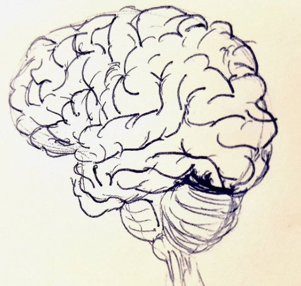
During one of my classes for 1st year Bachelor in Psychology, a student told me that he was interested in behaviour, and he could not understand why he had to focus so much on the brain. To answer this question, I like to dive a bit into philosophy.
Many philosophers, in fact, have dealt with the causes of behaviour. Their speculations can be divided into 3 main perspectives that have influenced the development of modern psychology:
🔴 Mentalism [e.g., Aristotle]: a person's mind (psyche) is a separate identity responsible for behaviour. This perspective has influenced modern behavioural science.
🔴 Dualism [e.g., Descartes]: behaviour is controlled by 2 entities, a mind and a body, that interact with each other. The mind receives information about the body and the world from the brain, and then it gives directives to the brain.
🔴 Materialism [e.g., Darwin]: behaviour can be measured objectively and consciousness is created in terms of physical properties of the brain.
Many still think that neuroscience is too far from actual psychology, because of the excessive focus on biology. This ignores the key value that a deep scientific knowledge of the nervous system and the links to behaviour may have in understanding a patient's condition and treatment options available.
Here I introduce some key topics about neuroanatomy and brain development as first steps to familiarize with neuroscience and the links between brain and behaviour.
When we think about neuroscience, most of us thinks about the brain. While the brain is key, it is not just about that!
The Central Nervous System is composed of brain and spinal cord, and it is the most complicated system in the human body. In particular, the brain weights between 1.2kg and 1.6kg, which is about 2% of the total body weight. Yet, the brain receives 20% of cardiac output for an adequate level of oxygen and energy. The main role of CNS is to mediate behaviour and it does that through a very complicated web of connections. It contains, in fact, as many as 86 billions neurons connected in a very intricate and unique way. No wonder that it is so complicated!
The nerve fibers that radiate out, as well as neurons outside the brain and spinal cord, constitute the Peripheral Nervous System. The PNS plays a fundamental role in:
🔴 transmitting sensation to the brain.
🔴 producing movement (as often, but not always, instructed by the brain).
🔴 balancing internal function (arousing and calming down).
🔴 controlling the gut.
By 24 days after conception, the cylinder expands towards the front to start forming the actual brain. By day 28 we will have the initial three brain vescicles:
The embryo of vertebrates, as well as their primitive stage of the nervous system, look very much alike. The different stages of development determine the great differentiation between species. While initially you may not recognise a human embryo from that of a chicken, by 49 days after conception it begins to resemble a person. The brain then looks clearly human by 100 days (around 3 months), but it does not have the classic folds and sulci that we are used to see in mature brains. Around 200 days (7 months) after conception the brain of the foetus begins to fold and it looks like an adult human brain by the end of the 9th month. Yet, the cellular structure is still very different!
The same cells that form the ectoderm give origin to very different systems in our body. When the ectoderm folds into the neural tube (which will become our brain and spinal cord), it leaves a layer of cells above. These cells will form the epidermis of the skin, of the hair, and of some glands. Some cells are also released when the neural tube forming the nervous system separates from the upper layer of the ectoderm (neural crest). These cells will take very different roles as in the gut or as facial cartilage, or take part into the development of lungs and heart and more. Alterations in the differentiation of these very different and complex roles can lead to disorders like ectodermal dysplasia, which shows symptoms in the skin, bones and teeth of the affected individuals.
Approaching neuroanatomy, I like to start by providing directions to navigate the brain. That is because I think that by learning directions we can visualize the brain better, and have the right instruments for orientation when playing with a brain atlas, online or in stains, or when dissecting an actual brain in a course. Learning these directions and their reflection onto nomenclature is fundamental to locate its structures from different perspectives.
Three main hypothetical planes thought to transect the human body are used as a reference for spatial orientation in the central nervous system:
• Horizontal plane = Transversal ≈ Axial [right figure]
• Vertical planes:
Different nomenclatures are used to label the same directions within the brain:
Proximal = close to a point of reference (medial if referred to the midline of the body)
Distal = away from a point of reference (lateral if referred to the midline of the body)
Inferior / Ventral = below
Superior / Dorsal = above
Anterior / Rostral / Frontal = toward the front
Posterior / Caudal = toward the back
Ventral = Towards the abdomen
Dorsal = Towards the back
Rostral = Toward the front
Caudal = Toward the tail
There are terms that were originally defined based on four-legged animals. However, the human brain has rotated of 90 degrees compared to these animals when gaining the upright posture over evolution. These terms are still used sometimes in certain nomenclatures of brain areas, and they might lead to some confusion. For example, caudal (i.e., towards the tail) indicates areas in the back side of the brain. Can you think about where the “human tail” would be? This would lead to a mistake in thinking that the caudal brain areas are located in the lower part of the brain in humans, while caudal still refers to the back. Similarly, ventral (i.e., towards the belly) indicates areas in the lower part of the brain and not towards the front (where our belly is actually located). You can then imagine how confusing this all can be!
Nevertheless, anatomical areas of the brain often include these terminologies in their names to indicate their position in the brain. For example, if I mention the "dorsolateral prefrontal cortex" you may even not have the most remote idea about what this region is and its function, but you can already imagine that this is located towards the face (frontal) but more on one side (lateral) and in the upper part of the brain (dorsal).
The neocortex (the most external 6 layers surrounding the brain hemispheres) can be divided into a number of regions based on their primary functions. These regions are called lobes and are:
Some sources also include a fifth lobe as in the insula, which is the cortex that separates the temporal lobe from the parietal and the frontal lobe and is dedicated to visceral sensation and autonomic function.
Within the different lobes, different brain areas have been defined based on the different cell types of the brain tissue in that specific area (Brodmann's classification).
The corpus callosum may be damaged or cut for medical reasons. For instance, a special surgery that cuts the corpus callosum, which is rarely performed in the 21st century, is an emergency, last option used to reduce epileptic seizures by preventing propagation of the excess of activity between the two hemispheres. This is called split brain condition, which produces behavioral symptoms that give information about the lateralization of brain processes. For instance, patients may not be able to recognise or name an object held in the left hand!
However, be aware that functional lateralization is not black-and-white! The two hemispheres collaborate to achieve most behaviours and damage in one hemisphere may lead to a functional deficit but this does not imply that the other hemisphere is not involved in that function at all!
Furthermore, some more recent studies have also shown an intact consciousness of objects in split-brain patients, even for objects held in the left hand. This is because visual information may still be in different degrees transmitted between hemispheres through a subcortical route!
• caudate nucleus
• putamen: works together with the caudate, and they are called striatum
• globus pallidus: also works together with the putamen, and they are called lentiform nucelus
• subthalamic nucleus and substantia nigra: more distant from the other nuclei but functionally related to them for motor control.
Functionally, the basal ganglia play an important role in motor control, motivation, reward-based learning, and addiction. Damage to these areas can lead to serious motor and cognitive deficits. An example? Motor alterations characteristic of Parkinson’s or Huntington’s disease.
The amygdala also originates from a ‘ganglion’ and some people consider it part of basal ganglia. However, given its multiple functional connections to other brain areas, it is more often considered to be part of the so-called limbic system.
This system is named so after its position at the border between brain hemispheres and brainstem. The hippocampus, named so because of its shape like a seahorse, is also part of the limbic system together with its neighbour, the amygdala, and the cingulate cortex. This system plays a major role in emotion recognition and response, motivation, olfaction, memory, autonomic regulation and sexual behaviours. The amygdala is the major structure for emotional processing, while the hippocampus is involved in declarative memory. In particular, it is involved in formation of new memory and semantic/episodic memory, not consolidated, before reaching the anterior cingulate cortex. It also encodes emotional context with the input from amygdala. A key aspect is also the spatial specificity of neuron firing that plays a major role in spatial memory and navigation.
Ever heard of London taxi drivers having a larger hippocampus?
The nervous system is not only the brain!
When we think about neuroscience, most of us thinks about the brain. While the brain is key, it is not just about that!
The Central Nervous System is composed of brain and spinal cord, and it is the most complicated system in the human body. In particular, the brain weights between 1.2kg and 1.6kg, which is about 2% of the total body weight. Yet, the brain receives 20% of cardiac output for an adequate level of oxygen and energy. The main role of CNS is to mediate behaviour and it does that through a very complicated web of connections. It contains, in fact, as many as 86 billions neurons connected in a very intricate and unique way. No wonder that it is so complicated!
The nerve fibers that radiate out, as well as neurons outside the brain and spinal cord, constitute the Peripheral Nervous System. The PNS plays a fundamental role in:
🔴 transmitting sensation to the brain.
🔴 producing movement (as often, but not always, instructed by the brain).
🔴 balancing internal function (arousing and calming down).
🔴 controlling the gut.
From here on I will focus on the brain for simplicity. Yet, be aware that there are many other players in the team!
It all starts with when a sperm fertilizes an egg. Then the new human consists of just a single cell (the human zygote). This cell immediately starts to divide and by day 15 the new embryo looks more like a fried egg (embryonic disc).
A primitive neural tissue forms as the outermost layer of cells in the embryo by 18 days after conception, and this is called neural plate. This neural plate is key for brain formation and development, and it is made of 2 layers of cells: the ectoderm and the endoderm.
By day 19, the ectoderm folds to form the neural groove, and then curls to form a cylinder which starts to zip up by day 22. Closure is completed by day 23, and with it the so-called neural tube. This tube is considered the nursery of the rest of the central nervous system.
Brain development.
It all starts with when a sperm fertilizes an egg. Then the new human consists of just a single cell (the human zygote). This cell immediately starts to divide and by day 15 the new embryo looks more like a fried egg (embryonic disc).
A primitive neural tissue forms as the outermost layer of cells in the embryo by 18 days after conception, and this is called neural plate. This neural plate is key for brain formation and development, and it is made of 2 layers of cells: the ectoderm and the endoderm.
By day 19, the ectoderm folds to form the neural groove, and then curls to form a cylinder which starts to zip up by day 22. Closure is completed by day 23, and with it the so-called neural tube. This tube is considered the nursery of the rest of the central nervous system.
By 24 days after conception, the cylinder expands towards the front to start forming the actual brain. By day 28 we will have the initial three brain vescicles:
1) prosencephalon or forebrain;
2) mesencephalon or midbrain;
3) rhombencephalon or hindbrain.
The remaining neural tube forms the spinal cord.
A bit over 1 month after conception, the prosencephalon expands on each side to form the brain hemispheres (telencephalon) and what will become thalamus and hypothalamus (diencephalon), which in turns expands in an optic outgrow as the forerunner of retina and optic tract.
The embryo of vertebrates, as well as their primitive stage of the nervous system, look very much alike. The different stages of development determine the great differentiation between species. While initially you may not recognise a human embryo from that of a chicken, by 49 days after conception it begins to resemble a person. The brain then looks clearly human by 100 days (around 3 months), but it does not have the classic folds and sulci that we are used to see in mature brains. Around 200 days (7 months) after conception the brain of the foetus begins to fold and it looks like an adult human brain by the end of the 9th month. Yet, the cellular structure is still very different!
The same cells that form the ectoderm give origin to very different systems in our body. When the ectoderm folds into the neural tube (which will become our brain and spinal cord), it leaves a layer of cells above. These cells will form the epidermis of the skin, of the hair, and of some glands. Some cells are also released when the neural tube forming the nervous system separates from the upper layer of the ectoderm (neural crest). These cells will take very different roles as in the gut or as facial cartilage, or take part into the development of lungs and heart and more. Alterations in the differentiation of these very different and complex roles can lead to disorders like ectodermal dysplasia, which shows symptoms in the skin, bones and teeth of the affected individuals.
How do we navigate the brain?
Three main hypothetical planes thought to transect the human body are used as a reference for spatial orientation in the central nervous system:
• Horizontal plane = Transversal ≈ Axial [right figure]
• Vertical planes:
- Coronal plane = Frontal plane [middle figure]
- Sagittal plane = Lateral plane (in the midline: Medial plane) [left figure]
Different nomenclatures are used to label the same directions within the brain:
Proximal = close to a point of reference (medial if referred to the midline of the body)
Distal = away from a point of reference (lateral if referred to the midline of the body)
Inferior / Ventral = below
Superior / Dorsal = above
Anterior / Rostral / Frontal = toward the front
Posterior / Caudal = toward the back
Ventral = Towards the abdomen
Dorsal = Towards the back
Rostral = Toward the front
Caudal = Toward the tail
There are terms that were originally defined based on four-legged animals. However, the human brain has rotated of 90 degrees compared to these animals when gaining the upright posture over evolution. These terms are still used sometimes in certain nomenclatures of brain areas, and they might lead to some confusion. For example, caudal (i.e., towards the tail) indicates areas in the back side of the brain. Can you think about where the “human tail” would be? This would lead to a mistake in thinking that the caudal brain areas are located in the lower part of the brain in humans, while caudal still refers to the back. Similarly, ventral (i.e., towards the belly) indicates areas in the lower part of the brain and not towards the front (where our belly is actually located). You can then imagine how confusing this all can be!
Nevertheless, anatomical areas of the brain often include these terminologies in their names to indicate their position in the brain. For example, if I mention the "dorsolateral prefrontal cortex" you may even not have the most remote idea about what this region is and its function, but you can already imagine that this is located towards the face (frontal) but more on one side (lateral) and in the upper part of the brain (dorsal).
Brain lobes.
The neocortex (the most external 6 layers surrounding the brain hemispheres) can be divided into a number of regions based on their primary functions. These regions are called lobes and are:
- Frontal lobe, dedicated to planning and reasoning.
- Parietal lobe, dedicated to processing and integration of sensory information.
- Occipital lobe, dedicated to processing of visual information.
- Temporal lobe, dedicated to emotion and memory.
Some sources also include a fifth lobe as in the insula, which is the cortex that separates the temporal lobe from the parietal and the frontal lobe and is dedicated to visceral sensation and autonomic function.
Within the different lobes, different brain areas have been defined based on the different cell types of the brain tissue in that specific area (Brodmann's classification).
Broadmann’s map of the cortex in humans and monkeys dates back to 1909! Many of the areas in Brodmann’s map have since been correlated closely to different brain functions. The idea was, in fact, that different cell types may be dedicated to different brain functions! Among most famous areas, we can mention Broca’s area (Broadmann’s area 44/45), involved in speech production, and Wernicke’s area (BA 22), involved in language comprehension. These areas are rather far apart in the brain (Broca’s in the frontal lobe, and Wernicke’s in the temporal lobe). This leads us to the concept of network. Although Broadmann’s studies shaped our understanding of brain anatomy, there is no perfect and simple match when mapping brain function to the specific structural areas defined by Broadmann. Clinical and modern neuroimaging studies have demonstrated, in fact, that complex brain functions are the result of the interaction of different brain areas in a network.
In terms of clinical implications, we can try to identify cortical areas that are possibly damaged in the case of certain behavioural symptoms. For instance, Broca’s area might be damaged when a person cannot speak fluently despite normal comprehension, while Wernicke’s area might be damaged if the person cannot understand the meaning of words.
In terms of clinical implications, we can try to identify cortical areas that are possibly damaged in the case of certain behavioural symptoms. For instance, Broca’s area might be damaged when a person cannot speak fluently despite normal comprehension, while Wernicke’s area might be damaged if the person cannot understand the meaning of words.
Structural connections.
White matter bundles, called commissures, are fibers that connect the two hemispheres. The largest commissure is the corpus callosum (containing 300 million fibers!), which is much more famous than the anterior commissure (the white matter bundles that connect left and right amygdala, and other medial temporal regions) and the posterior commissure (connecting nuclei involved in pupillary light reflexes).The corpus callosum may be damaged or cut for medical reasons. For instance, a special surgery that cuts the corpus callosum, which is rarely performed in the 21st century, is an emergency, last option used to reduce epileptic seizures by preventing propagation of the excess of activity between the two hemispheres. This is called split brain condition, which produces behavioral symptoms that give information about the lateralization of brain processes. For instance, patients may not be able to recognise or name an object held in the left hand!
However, be aware that functional lateralization is not black-and-white! The two hemispheres collaborate to achieve most behaviours and damage in one hemisphere may lead to a functional deficit but this does not imply that the other hemisphere is not involved in that function at all!
Furthermore, some more recent studies have also shown an intact consciousness of objects in split-brain patients, even for objects held in the left hand. This is because visual information may still be in different degrees transmitted between hemispheres through a subcortical route!
Deep in the brain!
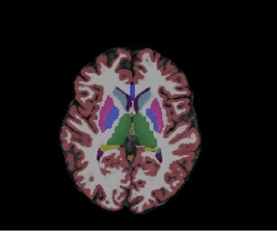
Finally, I’d like to focus on brain structures that are located deep in the brain, highly connected to the rest of the brain and supporting/regulating different key functions. These areas are more generally defined as subcortical areas, but we can also specify different subcortical systems.The basal ganglia are a group of connected nuclei forming a continuum at the base of the brain.
They are:
They are:
• caudate nucleus
• putamen: works together with the caudate, and they are called striatum
• globus pallidus: also works together with the putamen, and they are called lentiform nucelus
• subthalamic nucleus and substantia nigra: more distant from the other nuclei but functionally related to them for motor control.
Functionally, the basal ganglia play an important role in motor control, motivation, reward-based learning, and addiction. Damage to these areas can lead to serious motor and cognitive deficits. An example? Motor alterations characteristic of Parkinson’s or Huntington’s disease.
This system is named so after its position at the border between brain hemispheres and brainstem. The hippocampus, named so because of its shape like a seahorse, is also part of the limbic system together with its neighbour, the amygdala, and the cingulate cortex. This system plays a major role in emotion recognition and response, motivation, olfaction, memory, autonomic regulation and sexual behaviours. The amygdala is the major structure for emotional processing, while the hippocampus is involved in declarative memory. In particular, it is involved in formation of new memory and semantic/episodic memory, not consolidated, before reaching the anterior cingulate cortex. It also encodes emotional context with the input from amygdala. A key aspect is also the spatial specificity of neuron firing that plays a major role in spatial memory and navigation.
Ever heard of London taxi drivers having a larger hippocampus?


Comments
Post a Comment